Anatomy of lens:
The human eye contains a biconvex, transparent, crystalline structure called the lens, which is placed in-between iris and vitreous in the pattelar fossa. It plays an important role in the visual acuity.
It's refractive index is about 1.39 and total dioptric power is 18D. It's posterior surface is more convex than anterior & these surfaces meet at equator.
Structures of lens:
1) Lens capsule
- anterior lens capsule
- posterior lens capsule
2) Anterior epithelium
It is made of cuboidal epithelium. There is no posterior epithelium.
3) Lens fibres
These are the elongated epithelial cells.
4) Nucleus
It is the central part and it contains different zones, which corresponds to developmental periods.
a. Embryonic nucleus - 3 mth gestation
b. Fetal nucleus - 3mth gestation to till birth
c. Infantile nucleus - birth to puberty
d. Adult nucleus - after puberty.
5) Cortex
It is the peripheral part of nucleus consists of newly formed fibres.
Physiology of lens:
It is a transparent structure and it is important in focussing mechanism. It's physiological aspects include,
- lens transparency
- Metabolic activities of the lens
- Accomodation
Cataract:
Cataract means opacity in the lens (cataract-waterfalls). It is the most common condition affecting the vision.
Classification of cataract:
A) Based on etiology:
- Congenital & developmental cataract
- Acquired cataract
B) Based on morphology:
- Capsular (anterior & posterior)
- Subcapsular cataract (anterior & posterior)
- Cortical
- Supranuclear
- Nuclear
- Polar
Congenital & developmental cataracts:
It occurs due disturbance in the normal development of lens. In Congenital type cataract is limited to embryonic of foetal nucleus. Developmental cataract is limited to infantile or adult nucleus.
Etiology:
- Idiopathic
- Hereditary
- Maternal causes, malnutrition, infections,drugs ingestion, radiation.
- Foetal causes, anoxia, trauma, metabolic disorders, malnutrition.
Clinical Types:
1) Congenital capsular cataract
Rare condition and it occurs due to hyaloid artery remnants. It has anterior and posterior types.
2) Polar cataract
It occurs due to corneal perforation and delayed growth of anterior chamber. It occurs in center part of anterior capsule. It is also divided into anterior and posterior types.
3) Congenital nuclear cataract
- Cataracts pulverulenta
It is a bilateral condition occuring due to disturbance in lens development in very early stages. It appears exactly in the centre of lens.
- lamellar
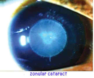
It is also called zonular cataract, where the opacity presents as discrete zones in the lens. It occurs due to Vit D deficiency, hypocalcemia and maternal rubella. It occurs in the zone of foetal nucleus surrounding the embryonic nucleus.
- sutural and axial
It consists of punctata opacities scattered around the Y sutures.
- total nuclear
It is characterized by bilateral chalky white dense opacity causes impairment of vision.It is non progressive.
4) Generalized cataract
- coronary
It is the most common form of developmental cataract occurs in puberty. It involves adult nucleus or deeper layer of cortex. The opacities are scattered and encircling the central axis. It does not affect vision until late stages.
- blue dot
It occurs in early two decades of life. It is also called cataracta punctata caerulea. It consists of punctata opacities as bluish dots in adult nucleus. It also doesn't affect vision when the opacity is small.
- total congenital
It occurs due to hereditary or maternal rubella.
Rubella cataract:
Maternal rubella infection during first trimester causes rubella cataract.
lens remains soft or even liquify. Cataractous nucleus may harbours the virus for two years.
- congenital membraneous cataract
There may be total or partial absorption of cataract leaving thin membraneous cataract. It is found in Hallermann Streiff Francois syndrome.


Social Plugin